|
Sino Biological
anti her2 car expression 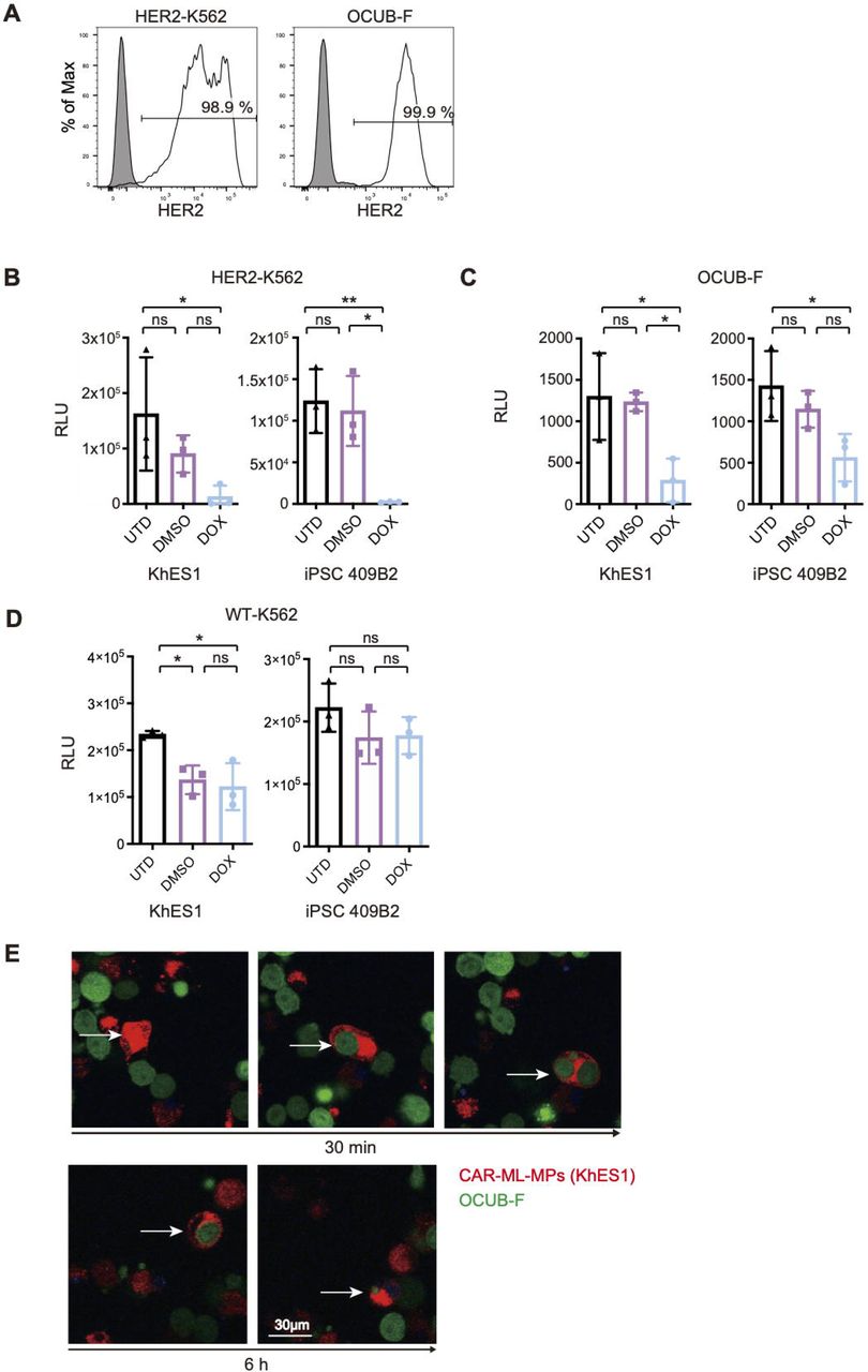 Anti Her2 Car Expression, supplied by Sino Biological, used in various techniques. Bioz Stars score: 94/100, based on 1 PubMed citations. ZERO BIAS - scores, article reviews, protocol conditions and more https://www.bioz.com/result/anti her2 car expression/product/Sino Biological Average 94 stars, based on 1 article reviews
anti her2 car expression - by Bioz Stars,
2026-03
94/100 stars
|
Buy from Supplier |
|
Cell Signaling Technology Inc
anti her2 antibody Anti Her2 Antibody, supplied by Cell Signaling Technology Inc, used in various techniques. Bioz Stars score: 96/100, based on 1 PubMed citations. ZERO BIAS - scores, article reviews, protocol conditions and more https://www.bioz.com/result/anti her2 antibody/product/Cell Signaling Technology Inc Average 96 stars, based on 1 article reviews
anti her2 antibody - by Bioz Stars,
2026-03
96/100 stars
|
Buy from Supplier |
|
Cell Signaling Technology Inc
her2 erbb2 rabbit mab Her2 Erbb2 Rabbit Mab, supplied by Cell Signaling Technology Inc, used in various techniques. Bioz Stars score: 96/100, based on 1 PubMed citations. ZERO BIAS - scores, article reviews, protocol conditions and more https://www.bioz.com/result/her2 erbb2 rabbit mab/product/Cell Signaling Technology Inc Average 96 stars, based on 1 article reviews
her2 erbb2 rabbit mab - by Bioz Stars,
2026-03
96/100 stars
|
Buy from Supplier |
|
Cell Signaling Technology Inc
phospho erbb2 tyr1221 1222 Phospho Erbb2 Tyr1221 1222, supplied by Cell Signaling Technology Inc, used in various techniques. Bioz Stars score: 96/100, based on 1 PubMed citations. ZERO BIAS - scores, article reviews, protocol conditions and more https://www.bioz.com/result/phospho erbb2 tyr1221 1222/product/Cell Signaling Technology Inc Average 96 stars, based on 1 article reviews
phospho erbb2 tyr1221 1222 - by Bioz Stars,
2026-03
96/100 stars
|
Buy from Supplier |
|
Cell Signaling Technology Inc
erbb2 her2 antibody Erbb2 Her2 Antibody, supplied by Cell Signaling Technology Inc, used in various techniques. Bioz Stars score: 95/100, based on 1 PubMed citations. ZERO BIAS - scores, article reviews, protocol conditions and more https://www.bioz.com/result/erbb2 her2 antibody/product/Cell Signaling Technology Inc Average 95 stars, based on 1 article reviews
erbb2 her2 antibody - by Bioz Stars,
2026-03
95/100 stars
|
Buy from Supplier |
|
Cell Signaling Technology Inc
p akt P Akt, supplied by Cell Signaling Technology Inc, used in various techniques. Bioz Stars score: 94/100, based on 1 PubMed citations. ZERO BIAS - scores, article reviews, protocol conditions and more https://www.bioz.com/result/p akt/product/Cell Signaling Technology Inc Average 94 stars, based on 1 article reviews
p akt - by Bioz Stars,
2026-03
94/100 stars
|
Buy from Supplier |
|
Cell Signaling Technology Inc
her2 erbb2 d8f12 xptm rabbit monoclonal antibody Her2 Erbb2 D8f12 Xptm Rabbit Monoclonal Antibody, supplied by Cell Signaling Technology Inc, used in various techniques. Bioz Stars score: 94/100, based on 1 PubMed citations. ZERO BIAS - scores, article reviews, protocol conditions and more https://www.bioz.com/result/her2 erbb2 d8f12 xptm rabbit monoclonal antibody/product/Cell Signaling Technology Inc Average 94 stars, based on 1 article reviews
her2 erbb2 d8f12 xptm rabbit monoclonal antibody - by Bioz Stars,
2026-03
94/100 stars
|
Buy from Supplier |
|
Cell Signaling Technology Inc
rabbit her2 erbb2 monoclonal antibody pe conjugate Rabbit Her2 Erbb2 Monoclonal Antibody Pe Conjugate, supplied by Cell Signaling Technology Inc, used in various techniques. Bioz Stars score: 93/100, based on 1 PubMed citations. ZERO BIAS - scores, article reviews, protocol conditions and more https://www.bioz.com/result/rabbit her2 erbb2 monoclonal antibody pe conjugate/product/Cell Signaling Technology Inc Average 93 stars, based on 1 article reviews
rabbit her2 erbb2 monoclonal antibody pe conjugate - by Bioz Stars,
2026-03
93/100 stars
|
Buy from Supplier |
|
Sino Biological
fitc anti her2 antibody  Fitc Anti Her2 Antibody, supplied by Sino Biological, used in various techniques. Bioz Stars score: 93/100, based on 1 PubMed citations. ZERO BIAS - scores, article reviews, protocol conditions and more https://www.bioz.com/result/fitc anti her2 antibody/product/Sino Biological Average 93 stars, based on 1 article reviews
fitc anti her2 antibody - by Bioz Stars,
2026-03
93/100 stars
|
Buy from Supplier |
|
Sino Biological
hrp conjugated rabbit anti her2 antibody  Hrp Conjugated Rabbit Anti Her2 Antibody, supplied by Sino Biological, used in various techniques. Bioz Stars score: 90/100, based on 1 PubMed citations. ZERO BIAS - scores, article reviews, protocol conditions and more https://www.bioz.com/result/hrp conjugated rabbit anti her2 antibody/product/Sino Biological Average 90 stars, based on 1 article reviews
hrp conjugated rabbit anti her2 antibody - by Bioz Stars,
2026-03
90/100 stars
|
Buy from Supplier |
|
Sino Biological
her2 erbb2 monoclonal antibody Her2 Erbb2 Monoclonal Antibody, supplied by Sino Biological, used in various techniques. Bioz Stars score: 94/100, based on 1 PubMed citations. ZERO BIAS - scores, article reviews, protocol conditions and more https://www.bioz.com/result/her2 erbb2 monoclonal antibody/product/Sino Biological Average 94 stars, based on 1 article reviews
her2 erbb2 monoclonal antibody - by Bioz Stars,
2026-03
94/100 stars
|
Buy from Supplier |
|
Sino Biological
her2 erbb2 cd340 antibody rabbit mab Her2 Erbb2 Cd340 Antibody Rabbit Mab, supplied by Sino Biological, used in various techniques. Bioz Stars score: 94/100, based on 1 PubMed citations. ZERO BIAS - scores, article reviews, protocol conditions and more https://www.bioz.com/result/her2 erbb2 cd340 antibody rabbit mab/product/Sino Biological Average 94 stars, based on 1 article reviews
her2 erbb2 cd340 antibody rabbit mab - by Bioz Stars,
2026-03
94/100 stars
|
Buy from Supplier |
Image Search Results
Journal: bioRxiv
Article Title: Enhanced anti-tumor effects through continuous administration of engineered CAR-macrophages derived from pluripotent stem cell-derived myeloid cell lines
doi: 10.1101/2024.07.22.604686
Figure Lengend Snippet: (A) Representative histograms showing HER2 expression in HER2-K562 and OCUB-F. (B, C) Cytotoxicity assay of CAR-ML-MPs derived from KhES1 and iPSC 409B2 strains against HER2-K562 (B) and OCUB-F (C). Effector-to-target ratio were 5:1 to HER2-K562 and 3:1 to OCUB-F, respectively (n = 3, independent experiment). UTD indicates untreated group. DMSO and DOX represent conditions treated without and with induction of CAR expression, respectively. RLU: relative light unit. Dunnett’s tests were performed as an a priori multiple comparison. *P < 0.05, **P < 0.01. Error bars represent mean ± standard deviation (SD). (D) Cytotoxicity assay of CAR-ML-MPs against wild type K562. (E) Results of time-lapse imaging of co-culture of CAR-ML-MPs (KhES1 strain) and OCUB-F (E/T ratio 1:1) after 30 minutes and 6 hours. Representative ML-MP cell (white arrow) showed phagocytosis of OCUB-F. See Video S1.
Article Snippet: For confirmation of
Techniques: Expressing, Cytotoxicity Assay, Derivative Assay, Comparison, Standard Deviation, Imaging, Co-Culture Assay

Journal: bioRxiv
Article Title: Enhanced anti-tumor effects through continuous administration of engineered CAR-macrophages derived from pluripotent stem cell-derived myeloid cell lines
doi: 10.1101/2024.07.22.604686
Figure Lengend Snippet: (A) Experimental time course of in vivo assays comparing single and consecutive tumor treatments. (B) Bioluminescence images and (C) quantitative values of bioluminescence from Luc-HER2-K562 cells at the indicated days and conditions. Statistical analysis was performed using a priori Dunnett’s multiple comparisons test. UTD indicates untreated group; 1 and 1+6 indicate single treatment alone and with additional treatment, respectively. *P < 0.05, **P < 0.01. Error bars represent mean ± standard deviation (SD). (D) Kaplan–Meier plot showing the survival of each group (n = 3 for each group). Statistical analysis was performed with Log-rank (Mantel–Cox) test. (E) Experimental time course of repeated Luc-ML-MP transplantation assay. Bioluminescence images of (F) Luc-ML-MPs and (G) their quantification (n = 3). Error bars represent mean ± standard deviation (SD).
Article Snippet: For confirmation of
Techniques: In Vivo, Standard Deviation, Transplantation Assay

Journal: bioRxiv
Article Title: Enhanced anti-tumor effects through continuous administration of engineered CAR-macrophages derived from pluripotent stem cell-derived myeloid cell lines
doi: 10.1101/2024.07.22.604686
Figure Lengend Snippet: (A) Schematic diagram of DOX-inducible anti-HER2 CAR construct. The construct includes HER2 recognition domain, intracellular hinge, CD28 and CD3ζ. TRE: tetracycline-response element. EF1: EF1 Alpha Promoter. TetR: Tet Repressor. ITR: inverted terminal repeat. (B) Proliferation curve of PSC-MLs derived from CAR-PSCs. On the left is mean ± standard deviation (SD) data for one passaging culture (n = 3, independent experiment). On the right, growth curves of KhES1-derived MLs cultured for 60 days. (C) Morphology of ML-MPs. Phase-contrast images (Upper panels) and May–Giemsa staining images (Lower panels). (D, E) Representative histograms showing macrophage cell-surface markers on CAR-ML-MPs. Panels (D) and (E) depict data from iPSC 409B2 and KhES1 strains, respectively. The rightmost panel displays histograms showing anti-HER2 CAR expression on the surface of CAR-ML-MPs in the presence of DOX. Gray fills represent isotype controls in all panels. (F) Expression of OX40L (left two panels) and 4-1BBL (right two panels). The relative quantity (Rq) of RNA expressions was measured in ML-MPs derived from the iPSC 409B2 strain (left) and the KhES1 strain (right) under both conditions, with and without DOX, compared to undifferentiated cells.
Article Snippet: For confirmation of
Techniques: Construct, Derivative Assay, Standard Deviation, Passaging, Cell Culture, Staining, Expressing

Journal: bioRxiv
Article Title: Enhanced anti-tumor effects through continuous administration of engineered CAR-macrophages derived from pluripotent stem cell-derived myeloid cell lines
doi: 10.1101/2024.07.22.604686
Figure Lengend Snippet: (A) Experimental time course of in vivo tumor treatment assay. (B, C) Bioluminescence images (B) and quantitative values of bioluminescence (C) from Luc-HER2-K562 cells at the indicated days and conditions. Statistical analysis was performed using a priori Dunnett’s multiple comparisons test. UTD indicates untreated group. DMSO and DOX represent conditions treated without and with induction of CAR expression, respectively. *P < 0.05, ***P < 0.001. Error bars represent mean ± standard deviation (SD). (D) Kaplan–Meier plot showing the survival of each group (n = 5 for each group). Statistical analysis was performed with Log-rank (Mantel-Cox) test. (E) Experimental time course of Luc-ML-MP transplantation assay. Bioluminescence images of (F) Luc-ML-MPs and (G) their quantification (n = 3). Error bars represent mean ± standard deviation (SD).
Article Snippet: For confirmation of
Techniques: In Vivo, Expressing, Standard Deviation, Transplantation Assay
Journal: Journal of Nanobiotechnology
Article Title: Dimeric Her2-specific affibody mediated cisplatin-loaded nanoparticles for tumor enhanced chemo-radiotherapy
doi: 10.1186/s12951-021-00885-6
Figure Lengend Snippet: Experimental workflow for the preparation of Pt@mPDA/MnO 2 /PDA-Z Her2 NPs and in vivo MRI-guided enhanced chemo-radiotherapy
Article Snippet: MCF-7 and SKOV-3 cells (2 × 10 5 ) were digested and resuspended in DMEM or McCoy’s 5A medium containing 2 or 4 μg/mL
Techniques: In Vivo

Journal: Journal of Nanobiotechnology
Article Title: Dimeric Her2-specific affibody mediated cisplatin-loaded nanoparticles for tumor enhanced chemo-radiotherapy
doi: 10.1186/s12951-021-00885-6
Figure Lengend Snippet: a SDS-PAGE of samples collected during Z Her2 preparation. M, protein marker; lane 1, total protein of uninduced cells; lane 2, total protein of induced cells; lane 3, soluble protein of induced cells; lane 4, insoluble protein of induced cells; lane 5, flow-through fractions after binding to Ni–NTA resin; lane 6, protein purified by Ni–NTA affinity chromatography. b SDS-PAGE of the purified Z Her2 affibody in the presence or absence of 2-ME. c SDS-PAGE characterization of Z Her2 labelled with 6-FAM. M, protein marker; lane 1, unlabelled Z Her2 ; lane 2, 6-FAM-labelled Z Her2 . d HAADF-STEM images of Pt@mPDA/MnO 2 /PDA-Z Her2 NPs. Element mapping of Pt@mPDA/MnO 2 /PDA-Z Her2 NPs: e merged image, f C, g N, h O, i S, j Mn, and k Pt
Article Snippet: MCF-7 and SKOV-3 cells (2 × 10 5 ) were digested and resuspended in DMEM or McCoy’s 5A medium containing 2 or 4 μg/mL
Techniques: SDS Page, Marker, Binding Assay, Purification, Affinity Chromatography

Journal: Journal of Nanobiotechnology
Article Title: Dimeric Her2-specific affibody mediated cisplatin-loaded nanoparticles for tumor enhanced chemo-radiotherapy
doi: 10.1186/s12951-021-00885-6
Figure Lengend Snippet: a CLSM images of SKOV-3 cells after 2 h incubation with Cy5.5@mPDA/MnO 2 /PDA NPs and Cy5.5@mPDA/MnO 2 /PDA-Z Her2 NPs and after a 1 h pre-treatment with or without Z Her2 . b Flow cytometry data for untreated SKOV-3 cells, SKOV-3 cells incubated for 4 h with Cy5.5@mPDA/MnO 2 /PDA NPs or Cy5.5@mPDA/MnO 2 /PDA-Z Her2 NPs, and cells pre-incubated with Z Her2 for 1 h before being exposed to Cy5.5@mPDA/MnO 2 /PDA-Z Her2 NPs for 4 h, and fluorescence intensity quantified. c MTT viability results for SKOV-3 cells after incubation with free cisplatin (Pt), Pt@mPDA/MnO 2 /PDA NPs, and Pt@mPDA/MnO 2 /PDA-Z Her2 NPs for 24 h. d Fluorescence images of calcein-AM/PI co-stained SKOV-3 cells after different treatments (dose of cisplatin: 48 μg/mL). e Intracellular ROS levels in SKOV-3 cells after treatment with different formulations
Article Snippet: MCF-7 and SKOV-3 cells (2 × 10 5 ) were digested and resuspended in DMEM or McCoy’s 5A medium containing 2 or 4 μg/mL
Techniques: Incubation, Flow Cytometry, Fluorescence, Staining
![a T 1 -MR images of SKOV-3 tumor-bearing mice before and after direct injection of Pt@mPDA/MnO 2 /PDA NPs (50 μL in PBS; [Mn] = 50 mM) into tumor (right, circle indicated) and muscle (left, circle indicated) tissues; b The quantified T 1 -MR signals corresponding to ( a ); c T 1 -weighted images of mice bearing SKOV-3 tumor grafts (arrow indicated) intravenously injection with Pt@mPDA/MnO 2 /PDA NPs or Pt@mPDA/MnO 2 /PDA-Z Her2 NPs ([Mn] = 3 mM, 100 μL in PBS per mouse) at 1 h, 3 h and 6 h, respectively; d The quantified T 1 -MR signals corresponding to ( c )](https://pub-med-central-images-cdn.bioz.com/pub_med_central_ids_ending_with_0847/pmc08120847/pmc08120847__12951_2021_885_Fig4_HTML.jpg)
Journal: Journal of Nanobiotechnology
Article Title: Dimeric Her2-specific affibody mediated cisplatin-loaded nanoparticles for tumor enhanced chemo-radiotherapy
doi: 10.1186/s12951-021-00885-6
Figure Lengend Snippet: a T 1 -MR images of SKOV-3 tumor-bearing mice before and after direct injection of Pt@mPDA/MnO 2 /PDA NPs (50 μL in PBS; [Mn] = 50 mM) into tumor (right, circle indicated) and muscle (left, circle indicated) tissues; b The quantified T 1 -MR signals corresponding to ( a ); c T 1 -weighted images of mice bearing SKOV-3 tumor grafts (arrow indicated) intravenously injection with Pt@mPDA/MnO 2 /PDA NPs or Pt@mPDA/MnO 2 /PDA-Z Her2 NPs ([Mn] = 3 mM, 100 μL in PBS per mouse) at 1 h, 3 h and 6 h, respectively; d The quantified T 1 -MR signals corresponding to ( c )
Article Snippet: MCF-7 and SKOV-3 cells (2 × 10 5 ) were digested and resuspended in DMEM or McCoy’s 5A medium containing 2 or 4 μg/mL
Techniques: Injection

Journal: Journal of Nanobiotechnology
Article Title: Dimeric Her2-specific affibody mediated cisplatin-loaded nanoparticles for tumor enhanced chemo-radiotherapy
doi: 10.1186/s12951-021-00885-6
Figure Lengend Snippet: Immunofluorescence assay of Cy5.5@mPDA/MnO 2 /PDA NPs and Cy5.5@mPDA/MnO 2 /PDA-Z Her2 NPs in SKOV-3 tumor tissues. Mice bearing SKOV-3 tumors were intravenously injected with Cy5.5@mPDA/MnO 2 /PDA NPs or Cy5.5@mPDA/MnO 2 /PDA-Z Her2 NPs (100 μL; 1 mg/mL in PBS). Tumors were collected and further studied by immunofluorescence assay after 12 h post-injection. a The immunofluorescence images of Cy5.5@mPDA/MnO 2 /PDA NPs (red) in tumor tissues; b The immunofluorescence images of Cy5.5@mPDA/MnO 2 /PDA-Z Her2 NPs (red) in tumor tissues; c Representative magnified immunofluorescence images corresponding to ( a ) and ( b ). Scale bar = 100 μm. The nuclei and Her2 were stained with DAPI (blue), anti-Her2 antibody (green), respectively
Article Snippet: MCF-7 and SKOV-3 cells (2 × 10 5 ) were digested and resuspended in DMEM or McCoy’s 5A medium containing 2 or 4 μg/mL
Techniques: Immunofluorescence, Injection, Staining

Journal: Journal of Nanobiotechnology
Article Title: Dimeric Her2-specific affibody mediated cisplatin-loaded nanoparticles for tumor enhanced chemo-radiotherapy
doi: 10.1186/s12951-021-00885-6
Figure Lengend Snippet: Bio-TEM images of Cy5.5@mPDA/MnO 2 /PDA NPs and Cy5.5@mPDA/MnO 2 /PDA-Z Her2 NPs in SKOV-3 tumor tissues. The red arrows indicate the NPs. Mice bearing SKOV-3 tumors were intravenously injected with Cy5.5@mPDA/MnO 2 /PDA NPs or Cy5.5@mPDA/MnO 2 /PDA-Z Her2 NPs (100 μL; 1 mg/mL in PBS). Tumors were collected and further studied by bio-TEM after 12 h post-injection
Article Snippet: MCF-7 and SKOV-3 cells (2 × 10 5 ) were digested and resuspended in DMEM or McCoy’s 5A medium containing 2 or 4 μg/mL
Techniques: Injection

Journal: Journal of Nanobiotechnology
Article Title: Dimeric Her2-specific affibody mediated cisplatin-loaded nanoparticles for tumor enhanced chemo-radiotherapy
doi: 10.1186/s12951-021-00885-6
Figure Lengend Snippet: In vivo chemotherapy efficacy of different formulations in mouse bearing SKOV-3 tumor grafts. When the volume of tumors reached about 50 mm 3 , mice were intravenously injected with cisplatin, Pt@mPDA/MnO 2 /PDA NPs or Pt@mPDA/MnO 2 /PDA-Z Her2 NPs (dose of cisplatin = 2 mg/kg) every 2 days. Tumor-bearing mice intravenously injected with the same volume of PBS were used as control group. The tumor volume and body weight were measured every day. On the 14 th day after inoculation, all the tumor grafts were removed, weighed and analyzed by TUNEL staining. a SKOV-3 tumor growth curves of different groups after intravenously injection of the formulations. b The images of tumors isolated after treatment for 8 days. c Mean weights of the tumors isolated on day 14. d Body weight changes over the 8 days of the experiments. e TUNEL-stained images of tumor slices excised from each treatment group on day 14. The nuclei of cells were visualized using DAPI (blue)
Article Snippet: MCF-7 and SKOV-3 cells (2 × 10 5 ) were digested and resuspended in DMEM or McCoy’s 5A medium containing 2 or 4 μg/mL
Techniques: In Vivo, Injection, TUNEL Assay, Staining, Isolation

Journal: Journal of Nanobiotechnology
Article Title: Dimeric Her2-specific affibody mediated cisplatin-loaded nanoparticles for tumor enhanced chemo-radiotherapy
doi: 10.1186/s12951-021-00885-6
Figure Lengend Snippet: In vivo chemo-radiation combined therapy efficacy of different formulations in mice bearing SKOV-3 tumor grafts. When the volume of tumors reached about 50 mm 3 , mice were intravenously injected with MnO 2 -free Pt@mPDA/PDA-Z Her2 NPs, Pt@mPDA/MnO 2 /PDA NPs or Pt@mPDA/MnO 2 /PDA-Z Her2 NPs every 2 days. Mice in control group were intravenously injected with same volume of PBS. Mice received an X-Ray radiation at a dose of 6 Gy for 24 h post-injection every 4 days except for PBS group. Tumor sizes and body weights were recorded every day. On the 14 th day after inoculation, all the tumor grafts were removed, weighed. a Schematic illustration of process of the chemo-radiation combined therapy; b SKOV-3 tumor growth curves of different groups after intravenously injection of the formulations; c The images of tumors isolated after treatment for 8 days; d Mean weights of the tumors isolated on day 14; e Body weight changes over the 8 days of the experiments
Article Snippet: MCF-7 and SKOV-3 cells (2 × 10 5 ) were digested and resuspended in DMEM or McCoy’s 5A medium containing 2 or 4 μg/mL
Techniques: In Vivo, Injection, Isolation
Journal: Journal of Nanobiotechnology
Article Title: Exosomes containing miRNAs targeting HER2 synthesis and engineered to adhere to HER2 on tumor cells surface exhibit enhanced antitumor activity
doi: 10.1186/s12951-020-00711-5
Figure Lengend Snippet: Downregulation of HER2 by miR-HER2-E1 and miR-HER2-E4 in SK-OV-3 and HEp-2 cells. a Immunoblotting analysis of HER2 protein levels of SK-OV-3 cells transfected with 0.5 μg of plasmids expressing miR-HER2-E1, miR-HER2-E4 or nontargeting (NT) miRNA. b The band densities of HER2 normalized to GAPDH indicating relative HER2/GAPDH expressing in SK-OV-3 cells were quantified and are presented as the mean ± standard deviation from three independent experiments. c HEp-2 cells were cotransfected with 0.5 μg of plasmid expressing miR-HER2-E1, miR-HER2-E4 or the NT miRNA and 0.2 μg of plasmid encoding His-tagged HER2. GAPDH served as a loading control. d The band densities of HER2 normalized to GAPDH indicating relative HER2/GAPDH expressing in HEp-2 cells were quantified and are presented as the mean ± standard deviation from three independent experiments. *p < 0.05
Article Snippet: After the coating solution was removed, nonspecific binding sites were blocked by incubation with 2% BSA at 37 °C for 1 h. The plates were rinsed, exposed to HER2 protein (Sino Biological, China) for 2 h, rinsed again, and reacted with the
Techniques: Western Blot, Transfection, Expressing, Standard Deviation, Plasmid Preparation, Control

Journal: Journal of Nanobiotechnology
Article Title: Exosomes containing miRNAs targeting HER2 synthesis and engineered to adhere to HER2 on tumor cells surface exhibit enhanced antitumor activity
doi: 10.1186/s12951-020-00711-5
Figure Lengend Snippet: Characterization of exosomes containing miR-HER2-E1. a Immunoblotting analysis of exosomes and cells with antibodies against the exosome marker proteins Alix, CD9, Annexin V, Flotillin-1 and TSG101. b The particle size distribution and number of isolated exosomes extracted from HEK-293 cells transfected with miR-HER2-E1 or non-targeting (NT) plasmid were measured by Izon’s qNano technology (Izon). c Quantification of miR-HER2-E1 from purified exosomes by qPCR analysis. The amount of exosomal miR-HER2-E1 was quantified and normalized to the amount of 18S rRNA. The data reported are representative of three independent experiments. All quantitative data are presented as the mean ± standard deviation
Article Snippet: After the coating solution was removed, nonspecific binding sites were blocked by incubation with 2% BSA at 37 °C for 1 h. The plates were rinsed, exposed to HER2 protein (Sino Biological, China) for 2 h, rinsed again, and reacted with the
Techniques: Western Blot, Marker, Isolation, Transfection, Plasmid Preparation, Purification, Standard Deviation

Journal: Journal of Nanobiotechnology
Article Title: Exosomes containing miRNAs targeting HER2 synthesis and engineered to adhere to HER2 on tumor cells surface exhibit enhanced antitumor activity
doi: 10.1186/s12951-020-00711-5
Figure Lengend Snippet: Inhibition of HER2 protein accumulation by exosome-delivered miR-HER2-E1. Immunoblotting analysis of HER2 protein levels in SK-OV-3 and HEp-2 cells treated with exosome-delivered miR-HER2-E1. a SK-OV-3 cells were left untreated (Con) or incubated with the indicated amounts of exosomes purified from HEK-293 cells transfected with miR-HER2-E1 or nontargeting (NT) miRNA. b The band intensity quantification shown at right represents the relative HER2/GAPDH expression levels in SK-OV-3 cell. c HEp-2 cells were transfected with the HER2 expression plasmid for 36 h and then either left untreated (Con) or incubated with 20 µg of purified exosomes produced by HEK-293 cells transfected with plasmids encoding the miR-HER2-E1 or the NT miRNAs. d The band intensity quantification shown at right represents the relative HER2/GAPDH expression levels in HEp-2 cells. The data reported are representative of three independent experiments. All quantitative data are presented as the mean ± standard deviation. *p < 0.05, N.S. indicates no significant difference
Article Snippet: After the coating solution was removed, nonspecific binding sites were blocked by incubation with 2% BSA at 37 °C for 1 h. The plates were rinsed, exposed to HER2 protein (Sino Biological, China) for 2 h, rinsed again, and reacted with the
Techniques: Inhibition, Western Blot, Incubation, Purification, Transfection, Expressing, Plasmid Preparation, Produced, Standard Deviation

Journal: Journal of Nanobiotechnology
Article Title: Exosomes containing miRNAs targeting HER2 synthesis and engineered to adhere to HER2 on tumor cells surface exhibit enhanced antitumor activity
doi: 10.1186/s12951-020-00711-5
Figure Lengend Snippet: miR-HER2-E1 delivered to HER2-positive cancer cells via exosomes reduces cell viability by activation of caspase-3/7. a Effects of exosome-delivered miR-HER2-E1 on the viability of HER2-positive (+) cancer cells (SK-OV-3 and HCT116) and HER2-negative (−) cells (HEp-2 and MDA-MB-231) measured by the CCK8 assay. Results are expressed as the mean of the cell viability index ± standard deviation compared to the mock-treated control (as 100%). b Similarly, the activities of caspase-3/7 were assessed by a Caspase‐Glo 3/7 assay kit after 24 h-treatment of exosomes. The caspase-3/7 activity is expressed as fold change compared to the mock treated (Mock) group. Data are presented as mean ± standard error of three independent experiments. Statistically significant differences between miR-HER2-E1 exosomes and the mock-treated are indicated by an asterisk (**p < 0.01; ***p < 0.001) in the HER2-positive (+) group. N.S. indicates no significant difference
Article Snippet: After the coating solution was removed, nonspecific binding sites were blocked by incubation with 2% BSA at 37 °C for 1 h. The plates were rinsed, exposed to HER2 protein (Sino Biological, China) for 2 h, rinsed again, and reacted with the
Techniques: Activation Assay, CCK-8 Assay, Standard Deviation, Control, Caspase-Glo Assay, Activity Assay

Journal: Journal of Nanobiotechnology
Article Title: Exosomes containing miRNAs targeting HER2 synthesis and engineered to adhere to HER2 on tumor cells surface exhibit enhanced antitumor activity
doi: 10.1186/s12951-020-00711-5
Figure Lengend Snippet: Antitumor efficacy of exosome-delivered miR-HER2-E1 in vivo. Nude mice bearing SK-OV-3 ( a ), HCT116 ( b ) or MDA-MB-231 ( c ) tumors (the average tumor size was 90 mm 3 for each group) were injected intratumorally every three days, 6 times in total (indicated by arrow), with 10 μg of purified exosomes per injection. Tumor size was measured every three days. Results are shown as the mean tumor volume (mm 3 ) ± standard deviation (n = 6). * and *** represent p < 0.05 and p < 0.001 compared with the NT exo group. N.S. indicates no significant difference
Article Snippet: After the coating solution was removed, nonspecific binding sites were blocked by incubation with 2% BSA at 37 °C for 1 h. The plates were rinsed, exposed to HER2 protein (Sino Biological, China) for 2 h, rinsed again, and reacted with the
Techniques: In Vivo, Injection, Purification, Standard Deviation

Journal: Journal of Nanobiotechnology
Article Title: Exosomes containing miRNAs targeting HER2 synthesis and engineered to adhere to HER2 on tumor cells surface exhibit enhanced antitumor activity
doi: 10.1186/s12951-020-00711-5
Figure Lengend Snippet: 293-miR-XS-HER2 expressing both a ligand for tumor cell surface HER2 and a miRNA targeting HER2. a Accumulation of miR-HER2-E1 in stable cell lines. miR-HER2-E1 isolated from 293-miR-XS and 293-miR-XS-HER2 cell pellets and exosomes was quantified with normalization to the level of 18S rRNA. b The relative HER2 binding affinity of 293-miR-XS and 293-miR-XS-HER2 exosomes . The absorbance readings (OD 450 nm) are shown on the Y axis. Each result is presented as the mean ± standard deviation of three replicates. **p < 0.01
Article Snippet: After the coating solution was removed, nonspecific binding sites were blocked by incubation with 2% BSA at 37 °C for 1 h. The plates were rinsed, exposed to HER2 protein (Sino Biological, China) for 2 h, rinsed again, and reacted with the
Techniques: Expressing, Stable Transfection, Isolation, Binding Assay, Standard Deviation

Journal: Journal of Nanobiotechnology
Article Title: Exosomes containing miRNAs targeting HER2 synthesis and engineered to adhere to HER2 on tumor cells surface exhibit enhanced antitumor activity
doi: 10.1186/s12951-020-00711-5
Figure Lengend Snippet: Antitumor efficacy of exosomes adhering to HER2 and expressing miR-HER2-E1. BALB/c-derived nude mice implanted with SK-OV-3 tumors with an average volume of 80 mm 3 were injected intravenously with exosomes purified from the parental HEK-293 cell line (293), the miR-HER2-E1-expressing stable cell line (293-miR-XS) or the stable cell line with coexpression of the HER2 protein ligand and miR-HER2-E1 (293-miR-XS-HER2). Exosomes were injected 3 μg/animal ( a ) or 30 μg/animal ( b ) every three days, for a total of 8 injections (indicated by arrow). The tumor size was measured every three days. The results are shown as mean tumor volume (mm 3 ) ± standard deviation (n = 6). * and *** represent p < 0.05 and p < 0.001 compared with the 293-miR-XS group
Article Snippet: After the coating solution was removed, nonspecific binding sites were blocked by incubation with 2% BSA at 37 °C for 1 h. The plates were rinsed, exposed to HER2 protein (Sino Biological, China) for 2 h, rinsed again, and reacted with the
Techniques: Expressing, Derivative Assay, Injection, Purification, Stable Transfection, Standard Deviation